Very often when a figure is wrong it easy to see without always knowing exactly what it is. That’s why it is important to have an idea of what is going on deep down under the surface of the skin to appreciate what’s happening close the surface.
On the skeleton there are places where the bones are very near to the overlying skin and these places are visible on most people, even those very overweight. Apart from many points on the head such as the forehead, cheekbones and chin, elsewhere there is also the wrist and anklebones amongst others. These areas are seen as depressions between the main muscle masses and sometimes as projections on thin people.
 |
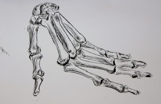
| Bones of the hand -conte crayon |
 |
| My own version |
After Andreas Vesaluiua De Humani Corporis Fabrica, 1543, New York Academy of Medicine from Drawing Lessons from theGreat Masters by Robert Beverly-Hale.
I knew from looking at this drawing that it is very complex. I didn’t intend to copy the whole figure at one go and soon after I started I began to realize just how complicated it is.
There are many muscles clustered around the top of the lower arm but each one can be broken down into just three groups: the supinator – rotates the hand, flexor - flexes and extensor – extends. The muscles in the hamstrings are also grouped in a similar way. The undulating lines both for the outlines and for those of the muscles really give a strong sense of movement. My own version certainly doesn’t live up to this description.
Rodin’s Seated Nude
There are many muscles clustered around the top of the lower arm but each one can be broken down into just three groups: the supinator – rotates the hand, flexor - flexes and extensor – extends. The muscles in the hamstrings are also grouped in a similar way. The undulating lines both for the outlines and for those of the muscles really give a strong sense of movement. My own version certainly doesn’t live up to this description.
Anatomy Sketch – from John Raynes Book
I found this drawing more enjoyable than the previous one. Some of
the shapes and angles were inaccurate in on my first attempt, so I adjusted these.
The most interesting part was doing the sketchy lines heading
in multiple directions and the way they flowed into one another.
On comparing this drawing with the life drawing I did of the standing
pose (one of the 3 Drawings exercises) it was easier to imagine much of the
underlying muscle structure.
|
Anatomy sketch Book illustration
|
Rodin’s Seated Nude
This to me is a good example of how flowing and undulating outlines make out the rounded forms as in that of the ribcage, the shapes of the major muscles and where the bones are close to the underlying skin.


No comments:
Post a Comment